
![]() Ultrasound
/ Interventional
Radiology
Ultrasound
/ Interventional
Radiology
![]() i
Current
pathology imaging
guidelines
i
Current
pathology imaging
guidelines
![]() @
Swiss
Physicians
email directory
@
Swiss
Physicians
email directory
Computed tomography
Also called CT-scan, "scanner" or simply CT, computed tomography has highly contributed, along with ultrasound, to the fantastic evolution of medical diagnosis in the seventies. Images reformatting from complex calculations has been made possible by a new generation of powerful computers.
Principles:
|
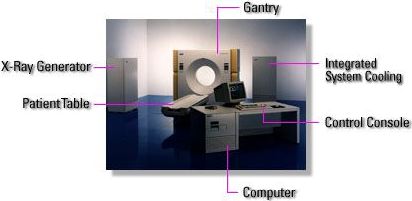
CT is based on the fact that X-ray absorption is proportional to density of the structures they go through to obtain a density profile of a body slice (transverse or axial cut).The patient is installed on a table moving horizontally inside a ring; an X-ray tube fixed to this ring generates a fan beam 1 to 10 mm thick rotating during 0.5-1s. around the patient; facing the tube are disposed thousands of detectors measuring the residual intensity of the beam transmitted through the body for each degree of rotation. |
 |
This slice information (raw data) is buffered in the computer, which then reconstructs an image composed of pixels after complex mathematic calculations. Each pixel is a tiny square of only a few tenths of a millimetre containing density information for a point in the slice. Densities are displayed as grey levels displaying the shape of body structures on an image looking finally like a print illustration.
Until beginning of the nineties, a CT examination was composed of separate slices, each slice position being defined by sequential progression of the moving table. Most today's scanners are also able to work in spiral mode, allowing a much faster scanning: the whole region is examined during a single breathhold lasting less than 30 seconds instead of obtaining separate breathhold slices. In spiral mode, the couple tube-detectors rotates continuously during the time necessary for the patient table to move through the gantry. Obtained data are submitted to additional calculations (interpolations) to obtain a 360 degree slice from a partial spiral (no more transverse) cut of the body. Spiral technique provides much more precise images less influenced by patient breathing; raw data may be then reconstructed in any space plane; surface or volume rendered images can also be computed.
Since a few years, an oustanding technology is boosting CT-scan use: multislice CT. Multiple parallel rows of detectors instead of only one are exposed to the X-ray beam, producing from 2 to 254 slices for one tube revolution. Examination time becomes very short (a few seconds) for a larger exploration field and very thin cuts. Due to volumic acquisitions instead of separate cuts, a huge quality improvement is obtained for native images as well as bi- or tridimensional reconstructions. The high speed thus obtained allows now for cardiac examinations. However, thousands of images are generated, imposing a heavier workload to the radiographer-radiologist team.
Dose-sparing techniques: recently, industry has designed iterative raw data reconstructions able to reduce the image background noise and obtain as much as 50% less radiation dose for the same result.
 |
|
|
Single slice Helical CT |
Mutlislice Helical CT |
Practically:
CT shows all deep-situated organs which are sometimes difficult to image by conventional X-rays or even ultrasound. CT is particularly useful for chest and abdomen diagnosis; an iodinated intravenous contrast medium is often required for better organ and blood vessels differentiation. CT is also often used to depict complex fractures. Angio-CT is obtained from millimetric slices performed after intravenous contrast injection to reconstruct arterial anatomy with a precision approaching arteriography. CT is usually contraindicated in pregnant women, as are most X-ray examinations.
 Specimen of chest and
abdominal CT images (with intravenous contrast enhancement)
Specimen of chest and
abdominal CT images (with intravenous contrast enhancement)
The radiologist has to perform continuous knowledge training, so that he will be be able to choose or advise the best suited imaging modality for every situation (X-rays, ultrasound, magnetic resonance, scintigraphy, etc.).
![]()