|
 Magnetic
Resonance
Magnetic
Resonance
 CT-Scan
 Digital
Radiology
Digital
Radiology
 Mammography
 Densitometry
 Ultrasound / Interventional
Radiology
Ultrasound / Interventional
Radiology
 i
Current
pathology imaging
guidelines i
Current
pathology imaging
guidelines
 @
Swiss
Physicians
email directory @
Swiss
Physicians
email directory
 Tips
page
Tips
page
 Links
Links
Abbreviations
The Hippocratic
Oath
Institute access map
Patient
prepararation
Site map
Home
Website in french
|
Approved by the "Fondation
Vaudoise
pour le Dépistage du cancer du sein"

Breast self-examination method
|

|
Breasts are submitted to continuous modifications
during
your menstrual cycle. A few days before bleeding,
women often notice
breast enlargment and mammary tension, which may even
be painful. That
is why you always have to do the self exam during the
same period of
your hormonal cycle, at best after your menstrual
period. At this time,
breast gland is usually softer and easier to feel.
Feeling is even more
efficient when the skin is wet, for example under the
shower or during
your bath. It is important to continue self exam
regularly after the
menopause. If you notice any breast change, do not
hesitate to show it
to your doctor, knowing that most of these phenomena,
for example
nodules or skin changes, are benign. For malignant
lesions, the sooner
a lesion is dectected, the better the outcome will be.
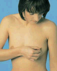
Images and information below
tell
you what you have to watch for and explain the
optimal way of doing
breast self exam in five steps.
|
1
The exam always begins with breasts inspection in the
mirror with your arms along your body. Look for
modifications of the
size of your breasts, of their shape, of the overlying
skin, surface
shrinkage/hump or deformation of the nipples. Cross
then your arms
above your head and repeat this operation.
|

|
|

|
2
In standing position, feel your Breasts with all the
fingers by flattening your hand; feel your right
breast with your left
hand and your left breast with your right hand.
|
3
It is a good method to begin palpation at the
internal
aspect of each breast and to progress towards the
external side.
Usually, the gland is denser in the upper external
part of the breast.
|

|
|
|
4
Examine separately the gland situated beneath the
nipple
as well as the nipple itself and the areola around it.
Gently squeeze
each nipple between your finger and thumb; if you see
nipple discharge,
notice its colour and its consistency and warn your
doctor.
|
5
Finally, feel your two armpits. Look for nodules.
Small
palpable lymph nodes are commonplace, but a
modification might be
important. Breast palpation must be repeated in
recumbent position,
because some abnormalities are only felt in this
position.
© AstraZeneca SA
Grafenau 10, 6301 Zoug
|

|

|